Determination of the morphological properties in the middle of the diaphysis of the third metatarsal bone of the crossbred horses
DOI:
https://doi.org/10.5281/zenodo.14103428Abstract
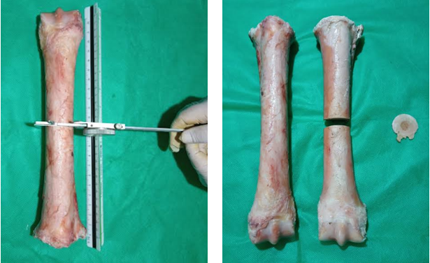
Within the complexity of equine diseases, musculoskeletal injuries are the most common cause of death, decline in performance and loss of training days. The morphological study of bone tissue reflects the importance of providing knowledge for this problem. The experimental determination of the morphological properties of bone tissue in horses is fundamental for animal clinics. This work aims to determine the morphological properties of horse metatarsal bones, providing knowledge and providing information with clinical purposes for the practice of Veterinary Medicine. The objectives of this work were: to determine the morphological characteristics of the metatarsal III of horses and to provide knowledge applicable to animal clinics. The metatarsals III of 20 Creole crossbred horses were studied, the weight and length of the bone were recorded. Transverse osteotomy was performed in the middle part of the diaphysis and the following measurements were made on the section surface: cortical thickness, cortical and medullary area. The variables under study were some for statistical analysis: descriptive, correlation, regression and variance with significance (p≤ 0.05). It is observed that there are significant differences between the areas (p< 0.0001). 87% of the total area in the middle part of the shaft of metatarsal III is occupied by the cortical area of the bone. The cortical area depends on the weight of the bone (R2= 0.88, p≤ 0.0001) and the age of the animals, being greater in G2 (p≤ 0.0014). The medial quadrant is the thickest, followed by the dorsal, lateral and plantar quadrants
Downloads
References
Agüera E. y Sandoval J. (1999). Anatomía Aplicada del Caballo. Harcourt Brace, España.
Baxter G. (2020). In Adams and Stashak’s Lameness in Horses, Seventh Edition.
Bigot G, Boudizi A., Rumelhart C. and Martin-Rosset W. (1996). Evolution during growth of the mechanical
properties of the cortical bone in equine cannon-bones. Medical Engineering and Physics. 18(1). 79-87.
Currey J. D. (1984). The mechanical properties of materials and the estructure of bone. In: The mechanical adaptation
of bone. University Press. Princeton. pp 3-37.
Espil J. I., Sánchez L., Dutra F., Guido N., López R., Barragán A., Alarcón F., Segura P., Quiroga M. A. (2022).
Fractura catastrófica condilar lateral del III hueso metatarsiano: reporte de caso en un equino pura sangre de
carrera del Hipódromo de La Plata. FAVE – Sección Ciencias Veterinarias 20 Suplemento Jornadas FCV-UNL
DOI:10.14409/favecv.2022.Suplemento.
Fioretti C., Galán A., Moine R., Varela M., Varela P., Mouguelar H., Gigena S., Bonino, F., Quinteros R., Natali J.
(2013). Características Mecánicas Dinámicas de la Tibia Aislada de Perro Sometida a Prueba de Impacto.
International Journal of Morphology. Chile. ISSN 0717 – 9502. 31 (2): 562-569.
Fioretti C., Natali J., Galán A., Rivera M., Moine R., Varela P., Varela M., Bonino F., Quinteros R. (2011).
Características Mecánicas Dinámicas del Fémur Aislado de Perro, Sometido Prueba de Impacto. International
Journal of Morphology. Chile. Vol. 29. 716:722.
Fioretti R., Moine R., Varela M., Varela P., Galán A., Gigena S., Mouguelar H., Gonzalez Sanchez S., Natali, J. (2018).
Densidad mineral ósea y resistencia ante la prueba de compresión en la mitad de la diáfisis del hueso fémur de
perro”. Revista Ab Intus. ISSN 1234-5678. Vol. 1, (1): 43-52.
Glade M, Belling T. (1984). Growth plate cartilage metabolism, morphology and biomechanical composition in over
and underfed horses. Growth. 48: 473.
Köning H, Liebich H. (2020). Veterinary Anatomy of domestic animals. Textbook and Colour Atlas. Tomo seventh
edition.Ed. Thieme. Alemania.
Medina L., Velásquez C.A., Figoli, M. (2011).Tratamiento de fractura transversa del hueso tercer metatarsiano en un
potrillo mediante la técnica de fijación interna. Revista Científica, vol. XXI, núm. 2, Universidad del Zulia
Maracaibo, Venezuela pp. 118-124.
Moine R., Galán M., Vivas A., Fioretti C., Varela M., Bonino F., Quinteros R., Natali J. (2015). Propiedades
Morfológicas en la Parte Media de la Diáfisis del Hueso Metacarpiano III de Equino Mestizo Criollo.
International Journal of Morphology Chile. ISSN 0717 – 9502. 33 (3): 955-961.
Moine, R., Rivera, C., Vivas, A., Ferraris, G., Galan, A., Natali, J. (2001): “Morfometría y determinación de calcio y
fósforo en la parte media de la diáfisis del metacarpiano III en yeguas mestiza con criollo”. Archivo Medicina
Veterinaria Chile. ISSN 0301 – 732X. XXXIII, N° 1, 63 – 68.
Moine R., Fioretti R.C., Galán A., Gigena S., Salvi M., Audap R., Varela M., Gonzalez Sanchez S., Varela P., Natali J.
(2020). Propiedades estructurales y resistencia a la flexión en tres puntos en la parte media de la diáfisis de la
falange proximal de la mano del caballo” Revista Ab Intus FAV-UNRC, 6 (3): 00-00 ISSN 2618-2734.
Natali J., Fioretti R., Moine R., Gigena S., Mouguelar H., Varela M, Varela P., Gonzalez Sanchez S., Quinteros R.,
Galán A. (2019) Morfología y comportamiento biomecánico de la falange proximal de la mano del caballo mestizo
criollo”. Revista Ab Intus. 56-62. ISSN 2618-2734. Vol. 3, (2): 43-52.
Nixon A. J. (2020). Equine Fracture Repair. Print ISBN:9780813815862 |Online ISBN:9781119108757.
Nunamaker D., Butterwerck D., Provost M. (1989). Some geometric of third metacarpal bone: a comparision between
the thoroughbred and standard bredrace horces. Journal of Veterinary Research, 22(2).129-134.
Southwood L. and Mclwaith C. (2000). Arthroscopic removal of abaxial fracture fragments involving a portion of the
base of the proximal sesamoid bone in horses. Journal of the American Veterinary Medical Association.
(2).236- 240.
Stover S. M. and Murray A. (2008). The California Postmortem Program: Leading the Way. Veterinary Clinics of
North America: Equine Practice. 24 (1): 21–36. https://doi.org/10.1016/j.cveq. 2007.11.009.
Yeni Y.N., Brown C.U., Wang Z. and Norman T. L. (1997). The influence of bone morphology on fracture toughness
of the human femur and tibia. Bone. 21 (5) pp 453- 459.
Published
How to Cite
Issue
Section
License
Copyright (c) 2024 Ab Intus

This work is licensed under a Creative Commons Attribution-NonCommercial 4.0 International License.