FETAL MUMMIFICATION: ALTERATION OF PLACENTAL COLLAGEN IN SOWS.
DOI:
https://doi.org/10.63207/ai.v8i15.162Keywords:
placenta, extracellular matrix, mummification, porcineAbstract
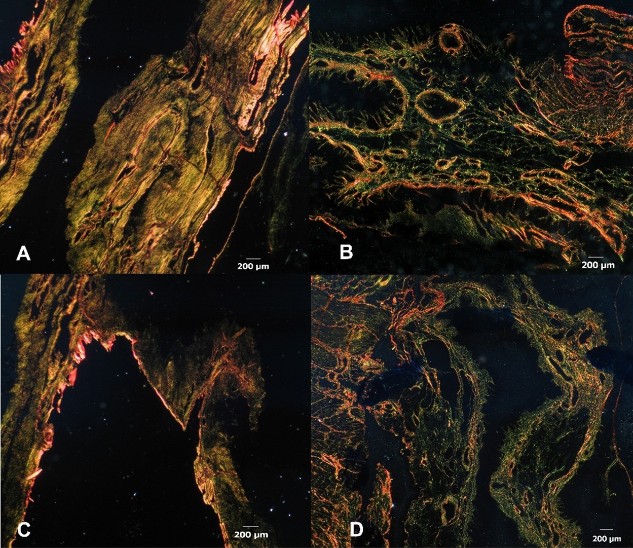
The objective was to determine the location of collagen in porcine placentas from mummified fetuses according to the thickness fiber. Placentas from crossbred sows from farms in the south of the province of Córdoba were collected after delivery and were processed. The fetoplacental units with mummified fetuses were selected and the gestational age was determined. Placentas of 60 and 90 days of pregnancy were collected. Collagen was evaluated through the Picrosirius-Polarization method. The arrangement of collagen fibers and the birefringence distribution were analyzed using the High Score value. The results were statistically analyzed and compared with previous studies carried in placentas from fetuses born alive. An association between the placental condition and the presence of fibers was determined. In placentas of mummified fetuses, a weak presence of fibers corresponding to collagen type I surrounded by large fibers of collagen type III were observed. An optimal placental microenvironment is essential for the development of physiological processes involved in maintaining pregnancy. An extracellular matrix with alterations in the composition and distribution of collagen would induce changes in the microenvironment incompatible with the normal development of gestation and fetal viability.
Downloads
References
Bonnans, C., Chou, J., Werb, Z. (2014). Remodelling the extracellular matrix in development and disease. Nature Reviews Molecular Cell Biology. 15(12): 786-801. doi: 10.1038/nrm3904
Borges, V., Bernardi, M., Bortolozzo, F., Wentz, I. (2005). Risk factors for stillbirth and foetal mummification in four Brazilian swine herds. Preventive Veterinary Medicine. 70: 165–176.
Cristofolini, A., Fiorimanti, M., Campos, M., Sanchis, E., Diaz, T., Moschetti E., Merkis, C. (2018). Morphometric study of the porcine placental vascularization. Reproduction in Domestic Animal. 53(1): 217-225. doi: 10.1111/rda.13095
Cury, D.P., Dias, F.J., Miglino, M.A., Watanabe I.S. (2016). Structural and ultrastructural characteristics of bone-tendon junction of the calcaneal tendon of adult and elderly Wistar rats. Public Library of Science One. 11(4): e0153568. doi:10.1371/journal. pone.0153568.
Di Rienzo, J., Casanoves, F., Balzarini, M., Gonzalez, L., Tablada, M., Robledo, C. (2020). InfoStat versión 2020. Argentina: Grupo InfoStat, FCA, Universidad Nacional de Córdoba.
Fernández, S.D., Amanto, F., Diéguez, S. (2019). Estrategias nutracéuticas que impactan sobre las pérdidas intrauterinas del feto porcino. Tesis de grado. Facultad de Ciencias Veterinarias. Universidad Nacional del Centro de la Provincia de Buenos Aires. Argentina. https://www.ridaa.unicen.edu.ar/handle/123456789/2122
Fiorimanti, M.R., Cristofolini, A.L., Gómez, K.P., Turiello, M.P., Bozzo, A.A., Barbeito, C.G., Merkis, C.I. (2024). Macroscopic and microscopic characterization of term placentas from nutritionally restricted goats. Small Ruminant Research. 233, 107232. https://doi.org/10.1016/j.smallrumres.2024.107232.
Fiorimanti, M.R., Rabaglino, M.B., Cristofolini, A.L., Merkis, C.I. (2018) Immunohistochemical determination of Ang-1, Ang-2 and Tie-2 I placentas of sows at 30, 60 and 114 days of gestation and validation through a bioinformatic approach. Animal Reproduction Science. 195: 242–250. https://doi.org/10.1016/j.anireprosci.2018.06.001
Gómez, K., Fiorimanti, M., Cristofolini, A., Benzoni, A.,Luján, M., Luján, O., Barbeito, C., Merkis, C. (2024). Caracterización del colágeno placentario fetal en cabras restringidas nutricionalmente durante la etapa prepuberal. Investigación Veterinaria. 26: 1-9. https://doi.org/10.62168/invet.v26i1.42
Johnson, G., Bazer, F., Seo, H. (2021). The early stages of implantation and placentation in the pig. Geisert, R.D and Spencer T.E (eds) In: Placentation in Mammals, Advances in Anatomy, Embriology and Cell Biology. 234: 61-90. https://doi.org/10.1007/978-3-030-77360-1_5
Liu, J., Xu, M.Y., Wu, J., Zhang, H., Yang, L., Lun, D.X., Hu, Y.C., Liu, B. (2021). Picrosirius-Polarization Method for Collagen Fiber Detection in Tendons: A Mini-Review. Orthopaedic Surgery. 13(3): 701-707. doi: 10.1111/os.12627.
Rittié, L. (2017). Method for picrosirius red-polarization detection of collagen fibers in tissue sections. Methods in Molecular Biology. 1627: 395-407.
Rozario, T., DeSimone, D.W. (2010). The extracellular matrix in development and morphogenesis: a dynamic view. Development Biology. 341(1): 126-40. doi: 10.1016/j.ydbio.2009.10.026.
Sanchis, E., Cristofolini, A., Taglialegna, A., Merkis, C. (2011). Moléculas de la matríz extracelular placentaria y uterina durante la preñez porcina. International Journal of Morphology. 29(4): 1438-1443.
Silva, G.S., da Costa Lana M.V., Días, G.B., da Cruz, R.A., Lopes, L.L., Machado, G., Corbellini, L.G., Gava, D., Souza, M.A., Pescador, C.A. (2015). Case-control study evaluating the sow’s risk factors associated with stillbirth piglets in Midwestern in Brazil. Tropical Animal Health and Production. 47(2): 445-9. doi: 10.1007/s11250-014-0745-8.
Soraci, A. 2012. Importancia práctica de los cambios fisiológicos en la alimentación del lechón. En UniRio Editora, Memorias XIº Congreso Nacional de Producción Porcina (CNPP11), las XVII Jornadas de Actualización Porcina (XVII JAP) y el VIº Congreso del Mercosur de Producción Porcina. Realizado en la ciudad de Salta entre 14 al 17 de agosto de 2012, Salta. Argentina. 43-50.
Trolliet, J. C. (2005). Productividad numérica de la cerda factores y componentes que la afectan. Disponible en: http://www.produccion-animal.com.ar/produccion_porcina/00-produccion_porcina_general/09-productividad_numerica_cerda.pdf
Van der Lende, T., Van Rens, D. (2003). Critical periods for fetal mortality in gilts identified by analising the length distribution of mummified fetuses and frecuency on non-fresh stillborn piglets. Animal Reproduction Science. 75:141-150.
Downloads
Published
How to Cite
Issue
Section
License
Copyright (c) 2025 Ab Intus

This work is licensed under a Creative Commons Attribution-NonCommercial 4.0 International License.